“Kafamız karıştı CIN nedir ? LSIL nedir? Doktorum” diyorsanız böyle buyrun lütfen 

“Kafamız karıştı CIN nedir ? LSIL nedir? Doktorum” diyorsanız böyle buyrun lütfen 

Servikal İntraepitelial Neoplazi Nedir? Nasıl Tedavi Edilip İzlenir?  (SIN / CIN  ya da LSIL /HSIL)

Aslında kafanızın karışması normal bizimki de çok karışmadı zamanında 🙂 🙂
 
Dolayısıyla kitaplarda “Terminoloji” başlığında anlatılan konuya hoş geldiniz.
 
Eskiden beridir rahim ağzının kanser öncülü (prekanseröz) lezyonlarını “Displazi” olarak adlandırıyoruz. Displaziler aslında “Servikal İntraepitelial Neoplazi” olarak tanımladığımız lezyonlar.
 
Yani nedir efendim:
“Servikal” yani rahim ağzında yer alan, “intraepitelial” yani epitel dediğimiz (ki cildimizi de benzer bir doku) dokunun içinde yer alan dışına taşmayan, “bazal membran” dediğimiz alt katmanı geçmeyen, “neoplazi” yani anormal gelişmiş dokulardır displaziler.
 
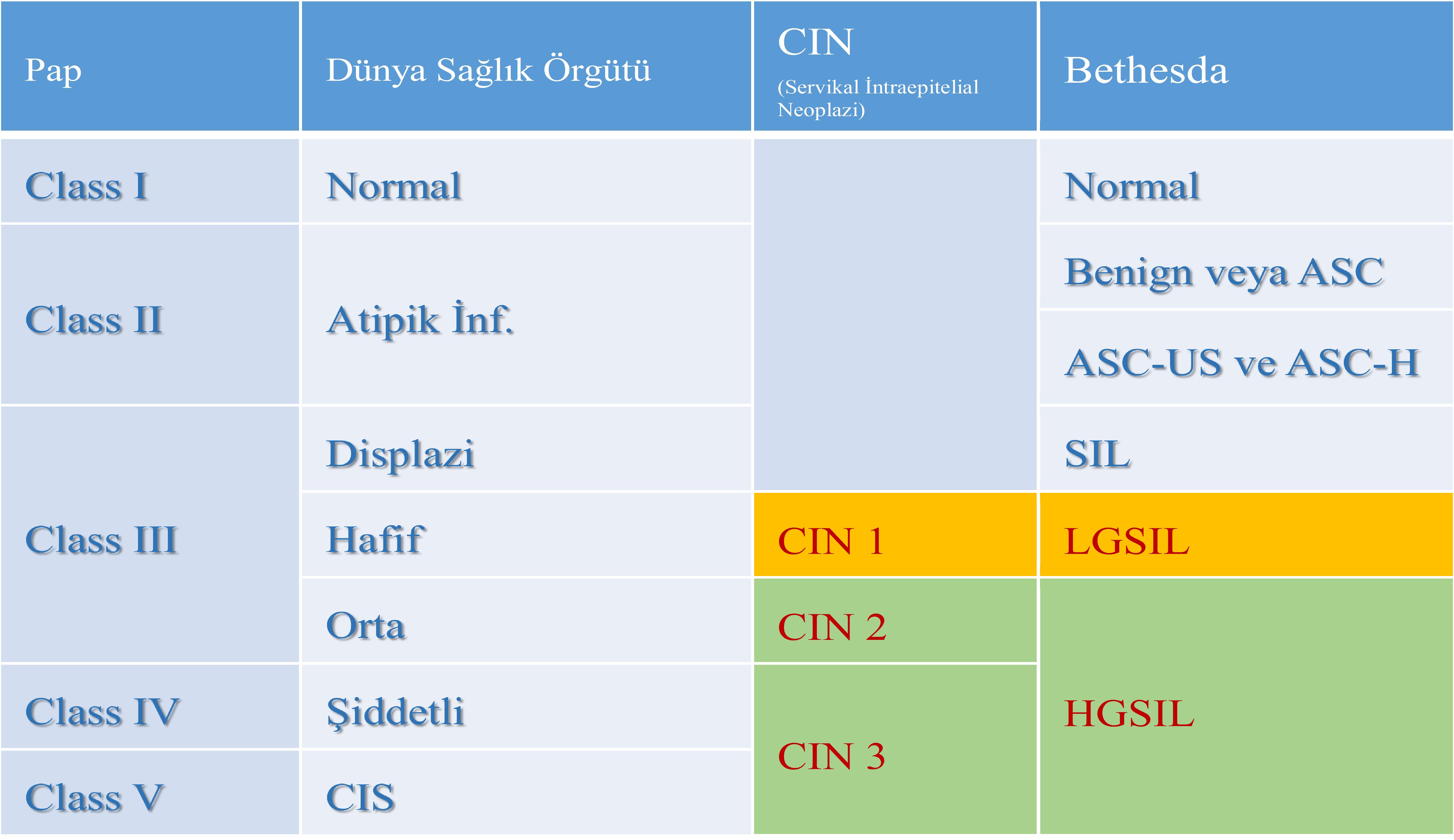
Displazileri de kendi içinde 3’e ayırıyorduk: Hafif (CIN I) Orta (CIN II) ve Ağır (CIN III)
 
Daha sonra 1988 yılında Amerikalılar biraz da literatürü kendileri domine etmek istedikleri için Bethesta sistemini getirdiler. Bu sınıflama çok mükemmel (!) olduğu için 1991 ve 2001 yıllarında 2 kez revize edildi.
 
Bu sistemede sitolojik hücrelerde (Smear de elde edilen hücrelerde) ve histolojik değerlendirmede (kolposkopi altında yapılan biyopsilerde) elde edilen hücrelerde farklı adlandırma yapıldı:
 
Pap smearde elde edilenler anormal hücreler için “Skuamöz intraepitelial neoplazi”, alınan biyopsilerden elde edilen hücreler için ise “servikal intraepitelial neoplazi” tanımlaması (Şu meşhur CIN olayı) kullanıldı.
 
Eee bana ne bu detaylardar derseniz. Sizi ilgilendiriyor zira elinize aldığınız patoloji raporunda mors alfabesi gibi yazılmış onlarca büyük harf kodlaması görüp sonra da “Google ana”nın kucağına atlıyorsunuz.
 
Okuduğunuz ve çoğunu anlamadığınız ki biz de bazılarını anlamıyoruz, tanımlamalardan sonuç çıkarmaya çalışırken sonunda “Evet ya kesin kanser oldum” noktasına varıp mebzul miktarda meslektaşımın ve benim telefonumu çaldırıyorsunuz.
 
İnanın çaldırmanız sorun değil de, bunca detayı telefonda açıklamaya çalışmak ama en önemlisi “kanser” olmadığınızı anlatmak çok zor oluyor. Dolayısıyla önemli 🙂 dostlarım. Ben anlatmaya, siz okumaya devam.
 
CIN’lerde kalmıştık. O zaman “Servikal İntraepitelial Neoplazi” (Cervical Intraepithelian Neoplasia) ne demek hücresel bağlamda dilimiz döndüğünce anlatalım:
 
Rahim ağzının dış kısmı, muayenede görülen kısmı skuamöz epitel ile kaplıdır. İşte bu dokunun en alt tabakası “bazal tabaka” diğer bir değiş ile temel tabakadır. Anormal hücreler, displazik hücreler, bazal tabakayı geçmedikleri sürece kanserden bahsedemeyiz! Bunu anlamanız önemli.
 
İşte bazal tabakanın üstünde kalan alanı patologlar kabaca üçe bölüyorlar.
 
CIN I: Bazal tabakanın üzerinde ki skuamöz epitel dokunun en alt, bazal tabakaya komşu 1/3’lük kısmında anormal, displazik hücrelerin bulunması durumudur.
 
CIN II’de ise skuamöz epitelin alt kısmından itibaren 2/3 kısmında displazik hücrelerin bulunmasıdır.
 
CIN III’de ise anlayacağınız gibi tüm epitelial katmanlarda anormal hücreler mevcuttur.
 
Bu noktada “Karsinoma in situ” tanımının üzerinde durmak gerek. Açıkçası “Karsinoma in situ” ile CIN III ya da HSIL arasında ki fark patologların arasında devamlı tartışılır.
 
Ve ne yalan söyleyeyim klinik bağlamda benim hastaya uygulayacağım tedaviyi değiştirmez. Ama klasik ekole bağlı ve sadece bu konu ile iştigal eden patologlar açısından “Karsinoma in situ” tanısı önemlidir ve ayırım mutlaka yapılmalıdır.
 
Örneğin değerli dostum patolog Prof.Dr.Ekrem Yavuz’un bu konuyu tartışırken “Ben kadın olsam karinoma in situ olup olmadığımı bilmek isterdim” dediğini gayet iyi hatırlıyorum.
 
Diğer taraftan şu terminoloji olayı da bitmek bilmedi.
 
2001 yılında Bethesta sistemi yenilendikten sonra bu sefer de 2012 yılında “Lower Anogenital Squamous Terminology (LAST)” projesi kapsamında CIN I’lere “Low Grade Squamous Intraepithelial Lesion = LSIL” denilmesine, CIN II ve III lezyonların ise “High Grade Squamous Intraepithelial Lesion = HSIL” olarak adlandırılmalarına karar verildi.
 
Ya yeter artık ne uğraşıyorlar derseniz valla ben de bilmiyorum ama konuştuğum yurdum topraklarında ki patoloji hocalarının anlattıkları onlarca öykü var.
 
Ama konu ile ilgili, benimde şahit olduğum en ilginç olaylardan biri 2010 yılında sadece HPV konuşulan “Eurogyn” toplantılarının birinde Fransız bir patolog meslektaşımızın kürsüye çıkıp “Yeter ben CIN II’mi geri istiyorum, size yedirmem” demesidir. O derece yani 🙂
(Not: Fransız profesör harbiden böyle dedi ben abartmadım ki abartmayı severim, çok ilginç ve komik gelmişti bize 🙂 )

Sonuç olarak aşağıda farklı sınıflamaları ve kullanılan terminolojileri tablo halinde görüyorsunuz.

Servikal Dispalzi Tablosu

29 Temmuz 2016 tarihinde Prof. Dr. Süleyman Engin Akhan tarafından yayınlanmış ve 27 Ağustos 2016 tarihinde de son güncelleme yapılmıştır.

Paylaş

Bir cevap yazın

E-posta hesabınız yayımlanmayacak. Gerekli alanlar * ile işaretlenmişlerdir

If you say “We are confused Doctor what is CIN ? What is LSIL?”, come to this section 

If you say “We are confused Doctor  what is CIN ? What is LSIL?”, come to this section 

What Is Cervical Intraepithelial Neoplasia? How Is It Treated and Monitored? (SIN / CIN or LSIL /HSIL))

In fact it is normal that you are confused, we were also very much confused once upon a time 🙂 🙂
 
Therefore welcome to the subject explained under the title of “Terminology” in books.
 
We refer to cervical cancer precursor (precancerous) lesions as “Dysplasia” from of old. Dysplasia is actually lesion that we identify as “Cervical Intraepithelial Neoplasia”.
 
So what is it?
“Servikal” Dysplasia is “neoplasia” in other words abnormally developed tissues not passing through the lower layer that we call “basal membrane”, which does not overflow and is included within “intraepithelial” in other words within the tissue that we call epithelium (it is a tissue similar to our skin) included in the cervix, in other words “cervical”.
 
We divide dysplasia into three within itself: Mild (CIN I) Medium (CIN II) and Severe (CIN III)
 
Later Americans brought Bethesda system in 1988 as they wanted to dominate the literature. This classification was revised for 2 times in 1991 and 2001 as it was excellent (!).
 
In this system, different designations were made in the cells achieved in cytological cells (in cells achieved in smear) and histological evaluation (in biopsies made under colposcopy):
 
“Squamous introepithelial neoplasia” was used for abnormal cells achieved in pap smear, “cervical intraepithelial neoplasia” definition was (That famous CIN incident) used for cells achieved from the biopsy received.
 
If you say, I don’t care about these details. It cares you because you apply to “Google mother” as you see tens of large letter codes written like morse alphabet in pathology report you receive.
 
While you try to reach a conclusion from the definitions that you read and don’t understand most of them, some of which we also don’t understand, at the end, you reach to a point that “Yes, I am cancer” and you call my colleague in beyond measure or you call me.
 
Believe me, it’s not a problem if you go crazy, but it is very difficult to try to explain all these details on phone but most importantly to explain that you are not “cancer”. Therefore it is important 🙂 my friends. I continue to explain and you continue to read.
 
We were talking about CINs. Then, let’s explain what “Cervical Intraepithelian Neoplasia” means in cellular respect as much as I can:
 
Outer part of the cervix, the part seen in the examination, is covered with squamous epithelium. So the lowest layer of this tissue is “basal layer” in other words basic layer. As long as abnormal cells, dysplasia cells do not pass basal layer, we cannot talk about cancer! It is important that you understand it.
 
The area above basal layer is roughly divided into three by pathologists.
 
CIN I: It is the case of presence of abnormal dysplasia cells in 1/3 part neighboring the bottom basal layer of squamous epithelium tissue on basal layer.
 
It is the presence of dysplasia cells in 2/3 part as of lower layer of squamous epithelium in CIN II.
 
There are abnormal cells in all epithelial layers in CIN III as you can understand.
 
At this point, we should focus on “Carcinoma in situ” definition. Obviously the difference between CIN III or HSIL with “Carcinoma in situ” is always discussed among pathologists.
 
And I should say that it doesn’t clinically change the treatment I shall apply on the patient. But “Carcinoma in situ” diagnosis is important for pathologists, who are bound to classic ecole and only engaged in this issue, and differentiation must be made.
 
For instance; I remember very well that my valuable friend pathologist Prof.Dr.Ekrem Yavuz said “If I were a woman, I would know if I were carcinoma in situ.” while discussing this issue.
 
On the other hand, this terminology incident still continues.
 
After Bethesda system was renewed in 2001, this time in 2012 it was decided that CIN I was referred to as “Low Grade Squamous Intraepithelial Lesion = LSIL” and CIN II and III lesions were referred to as “High Grade Squamous Intraepithelial Lesion = HSIL” within the scope of “Lower Anogenital Squamous Terminology (LAST)” project.
 
If you say “It is enough. What are they trying for?”, I also don’t know but there are tens of stories explained by pathology masters that I talk to in my country.
 
However, one of the most interesting incidents that I witness related to the issue is that one of our French pathologist colleague went up to the rostrum in one of “Eurogyn” meetings, in which only HPV was talked, in 2001 and said, “Enough. I want my CIN II back, you cannot cancel it”. It is such an extent:-)
(Note: French professor told exactly the same, I didn’t exaggerate even though I like to exaggerate, it was very interesting and funny to us:-) )

As a result, you can see different classifications and terminologies used below as a table.

Servikal Dispalzi Tablosu

29 Temmuz 2016 tarihinde Prof. Dr. Süleyman Engin Akhan tarafından yayınlanmış ve 20 Kasım 2018 tarihinde de son güncelleme yapılmıştır.

Paylaş

Bir cevap yazın

E-posta hesabınız yayımlanmayacak. Gerekli alanlar * ile işaretlenmişlerdir